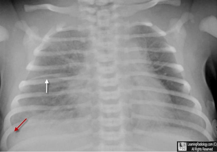
Baby Chest X Ray Pneumonia
In our patient with a pulmonary consolidation, the clinical findings of fever, cough and elevated wbc count raise infection (i.e. The inappropriate use of cxr results in children exposure to ionizing radiations and increased medical costs.

Pin On Radiology For Clinicians
Please see disclaimer on my website.

Baby chest x ray pneumonia. The differential for the radiologic finding of pulmonary consolidation includes blood (pulmonary hemorrhage), pus (infection, i.e. 7 if a child is dehydrated, especially early in the course of illness, an. • cough, tachypnea, hypoxia • asymmetric breath sounds, rales, decreased air movement • fever • elevated wbc with left shift
Appearance of gas filled loops in the chest may appear only few hours after birth. And the testing folder contains 234 normal and 390 pneumonia images. Uk—do not routinely perform a chest x ray in children with bronchiolitis because changes on x ray may mimic pneumonia and should not be used to determine the need for antibiotics.
This dataset was acquired from picture archiving and communication systems (pacs). Chest radiographs (cxrs) are the most widely employed test, however, they are not indicated in ambulatory settings, cannot distinguish between viral and bacterial infections and have a limited role in the ongoing management of disease. These findings are not specific to this pneumonia.
However, cxr is still performed in a high percentage of cases, mainly to diagnose or rule out pneumonia. Although a positive chest radiograph is the gold standard for the diagnosis of pneumonia, an occasional patient with clinical symptoms and signs of pneumonia may have a normal chest roentgenogram. Here we review the role of radiology in the diagnosis of paediatric pneumonia.
The validation folder, however, contains only 8. If one desires to delineate the Pneumonia) to the top of our differential.
A soft rubber tube is better than an infant feeding tube for the radiological diagnosis of tef. They belonged to a cohort examined in connection with the introduction of rapid methods for virological diagnosis. Morgagni hernias are seen as opacities adjacent to the right costophrenic angle.
Read more 1 doctor agrees Imaging should be restricted to children who appear toxic, those with the recurrent or prolonged course of illness despite. Both cases showed bilateral interstitial infiltrates and hyperinflation.
20 the radiographic finding is said to lag behind the clinical picture. 2 weeks of antibiotics, then another x. Browse 229 chest x ray pneumonia stock photos and images available or start a new search to explore more stock photos and images.
Chest radiography findings in children with tuberculous pneumonia may include hilar or mediastinal lymphadenopathy, atelectasis, or consolidation of a segment or lobe (usually right upper lobe), pleural effusion, cavitary lesions (in. Pneumonia), fluid (heart failure), and cells (cancer). Consider performing a chest x ray if intensive care is being proposed for a child.

Walking Pneumonia Walking Pneumonia In Children Pneumonia In Kids Health Info Lungs Health

Chest Xray Severe Changes From Chronic Tuberculosis Radiologist Radiology Cough This Information Is Protected Health Information Radiology Radiologist

Gpawegeners Granulomatosis - X-ray Case 190 Radiology X Ray Pathology

How Long Is My Loved One Going To Stay In Intensive Care With Pneumonia Find The Answer And Lots Of Free Resources At H Pneumonia Healthy Lungs Intensive Care

Pin On X-ray

Pneumonia In Children Intechopen Beautiful Bodies Pneumonia In Kids Beautiful

Pediatric Radiology Pediatric Radiology Radiology Pediatrics

Pin On Medicine

Ap Ribs- Used To Visualize Posterior Ribs X Ray Visual Radiology

Ttn Transient Tachypnea Of Newborn Radiology Bronchopulmonary Transients

Pin By Jennifer Larson On Extreme X-rays Radiology Humor Radiology Radiology Imaging

Chest Xray Of A Child With A Cough Shows Pneumonia Radiologist Radiology Diagnostic Imaging X Ray Medical Imaging

Pin On Nurse Practitioner

Learningradiology - Round Pneumonia Pediatric Radiology Pneumonia Radiology

The Radiology Assistant Chest X-ray - Lung Disease Lung Disease Medical Radiography Radiology Imaging

Achondroplasia Radiology Case Radiopaediaorg Achondroplasia Radiology

Chest Pa Digital X-ray Image Made By A Cr Unit

Dark Lung Fields Medical Radiography Radiology Cardiology Nursing

Chest Xray In A Child With Cough Shows Round Pneumonia Radiologist Radiology Radiology X Ray Radiologist